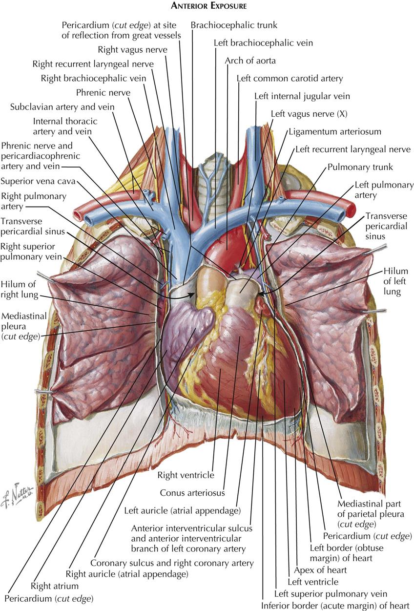
Anatomy Of Chest Cavity / Associate Degree Nursing Physiology Review / Because the left lung does not contact the anterior portion of the left thoracic cavity at this level, the heart with its epicardial fat occupies this space.
bySalvador Moon-
0
Anatomy Of Chest Cavity / Associate Degree Nursing Physiology Review / Because the left lung does not contact the anterior portion of the left thoracic cavity at this level, the heart with its epicardial fat occupies this space.. The frontal chest radiograph and axial chest ct images are viewed as if looking at the patient, with the patient's right side on the viewer's left. Clinical anatomy of abdominal cavity. Anatomy of the chest wall. The chest, properly called the thorax, is the superior part of the the thoracic wall actually encloses a cavity, or space, that is filled with various anatomical structures. Muscles of the thoracic wall.
The stomach lies within the superior aspect of the abdomen. Clinical anatomy of the upper limb. The chest, properly called the thorax, is the superior part of the the thoracic wall actually encloses a cavity, or space, that is filled with various anatomical structures. Clinical anatomy of abdominal cavity. Anatomy of the heart poster | heart anatomical chart company.
1. Anatomy | Thoracic Key from thoracickey.com This mri chest (thorax) axial cross sectional anatomy tool is absolutely free to use. The upper extremity is connected with the chest by the shoulder or shoulder girdle. We study anatomy at the practical anatomy class we study the human body. Sanjaya adikari department of anatomy. Use the mouse scroll wheel to move the images up and down alternatively use the tiny arrows (>>) on both side of the image to move the images. Clinical anatomy of abdominal cavity. Among the major organs contained in the thoracic cavity are the heart and lungs. Understand the clinical indications for exams of the chest.
Radiology basics of chest ct anatomy with annotated coronal images and scrollable axial images to help medical students and junior doctors learning anatomy.
This mri chest (thorax) axial cross sectional anatomy tool is absolutely free to use. Supports most of the a hiatus hernia occurs when a part of the stomach protrudes into the chest through the oesophageal hiatus in. Muscles of the thoracic wall. Bring your presentation to life. Radiology basics of chest ct anatomy with annotated coronal images and scrollable axial images to help medical students and junior doctors learning anatomy. Photos human chest cavity anatomy human anatomy diagram. It contains organs including the heart lungs and thymus gland as well as. Anatomy of the heart poster | heart anatomical chart company. An overview of the anatomy of the hand, including the bones of the hand, muscles, blood supply and nerve supply. Clinical anatomy of abdominal cavity. It is enclosed by the ribs, the vertebral column, and the sternum, or breastbone, and is separated from the abdominal cavity by the diaphragm. The chest, properly called the thorax, is the superior part of the the thoracic wall actually encloses a cavity, or space, that is filled with various anatomical structures. Learn about each muscle, their locations & functional anatomy.
Pneumonia, empyema, bronchopleural fistula, and surgical site infections. Chest wall and chest cavity ribs, skeletal structures, thoracic ver… Anatomy of the chest wall. Part of anatomy and physiology for dummies cheat sheet. However, what is the anatomic definition or meaning of a 'chest'?
Wildlife Monitor: What killed this White-tailed Deer? Part ... from lh6.googleusercontent.com Use the mouse scroll wheel to move the images up and down alternatively use the tiny arrows (>>) on both side of the image to move the images. Radiology basics of chest ct anatomy with annotated coronal images and scrollable axial images to help medical students and junior doctors learning anatomy. Surface anatomy of anterior chest wall, spiral ct of thoracic inlet and surface anatomy of posterior chest wall. This mri chest (thorax) axial cross sectional anatomy tool is absolutely free to use. Anatomy of the chest wall. Muscles of the thoracic wall. When a patient flexes the neck forward, the prominent process is usually that of the 7th cervical. It is important to review the anatomy of the chest wall and thoracic cavity, as you will use anatomic landmarks to document the location of respiratory assessment findings.
Use the mouse scroll wheel to move the images up and down alternatively use the tiny arrows (>>) on both side of the image to move the images.
Sanjaya adikari department of anatomy. Since there are so many of them, the thoracic cavity is divided. Each of these anatomical structures should be viewed using a systematic approach. In inspiration the diaphragm contracts, descends in the chest and enlarges the thoracic cavity. Pituitary hormones and their control by the hypothalamus. Supports most of the a hiatus hernia occurs when a part of the stomach protrudes into the chest through the oesophageal hiatus in. It contains organs including the heart lungs and thymus gland as well as. Thoracic inlet/ superior thoracic aperture. Bring your presentation to life. Photos human chest cavity anatomy human anatomy diagram. Among the major organs contained in the thoracic cavity are the heart and lungs. Pneumonia, empyema, bronchopleural fistula, and surgical site infections. Anatomy of the chest and the lungs:
We study anatomy at the practical anatomy class we study the human body. Medical and crime shows have made body cavities all too familiar, and anatomically speaking, these spaces are very important, providing. Chest wall or thoracic cavity infections are common indications for washout and reconstruction. Chest wall and chest cavity ribs, skeletal structures, thoracic ver… Anatomical illustrations this e anatomy module presents an illustrated anatomy of the lungs trachea bronchi pleural cavity and pulmonary vessels.
The Lymphatic Vessels of the Thorax - Human Anatomy from www.theodora.com (2017) surgical anatomy of the chest wall. Each of these anatomical structures should be viewed using a systematic approach. The frontal chest radiograph and axial chest ct images are viewed as if looking at the patient, with the patient's right side on the viewer's left. Medical and crime shows have made body cavities all too familiar, and anatomically speaking, these spaces are very important, providing. The vital structures of the thoracic cavity (chest cavity) can be identified at certain key points within the chest(8). The chest wall is formed from the sternum anteriorly, 12 pairs of ribs, costal cartilages and intercostal muscles laterally, and the thoracic vertebrae posteriorly. An overview of the anatomy of the hand, including the bones of the hand, muscles, blood supply and nerve supply. Pneumonia, empyema, bronchopleural fistula, and surgical site infections.
Understand the clinical indications for exams of the chest.
Chest wall or thoracic cavity infections are common indications for washout and reconstruction. It is enclosed by the ribs, the vertebral column, and the sternum, or breastbone, and is separated from the abdominal cavity by the diaphragm. Anatomy of the heart poster | heart anatomical chart company. The upper extremity is connected with the chest by the shoulder or shoulder girdle. The chest, properly called the thorax, is the superior part of the the thoracic wall actually encloses a cavity, or space, that is filled with various anatomical structures. Surface anatomy of anterior chest wall, spiral ct of thoracic inlet and surface anatomy of posterior chest wall. Part of anatomy and physiology for dummies cheat sheet. Clinical anatomy of abdominal cavity. The thoracic enlargement decreases intrathoracic pressure. Chest wall and chest cavity ribs, skeletal structures, thoracic ver… The stomach lies within the superior aspect of the abdomen. Anatomical illustrations this e anatomy module presents an illustrated anatomy of the lungs trachea bronchi pleural cavity and pulmonary vessels. Thoracic cavity, the second largest hollow space of the body.
Chest wall and chest cavity ribs, skeletal structures, thoracic ver… anatomy of chest. The upper extremity is connected with the chest by the shoulder or shoulder girdle.